
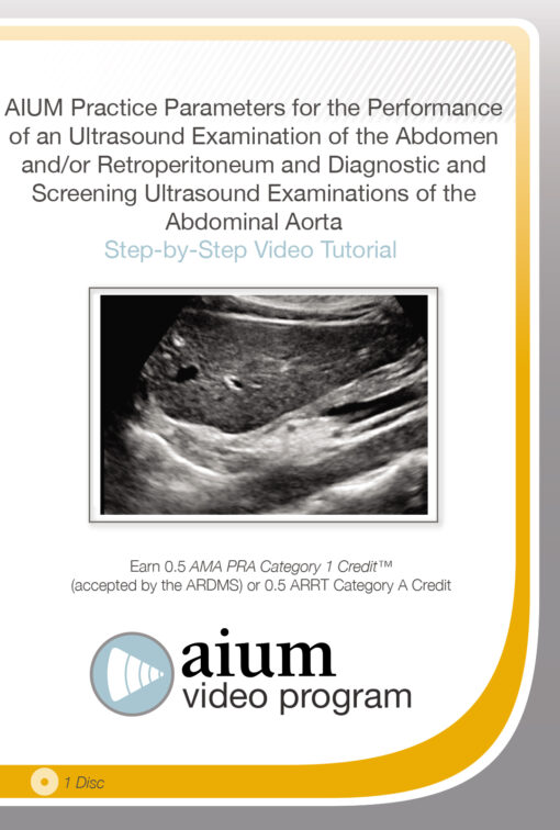
AIUM Practice Parameter for the Performance of an Ultrasound Examination of the Abdomen and/or Retroperitoneum and Diagnostic and Screening Ultrasound Examinations of the Abdominal Aorta
$18.00
This Product is shared via google drive download link, So please share your correct Gmail id while placing the order .Please note that there are no CME points or certificate associated with this course Samples for Courses Can be found here : Free Samples Here!
AIUM Practice Parameter for the Performance of an Ultrasound Examination of the Abdomen and/or Retroperitoneum and Diagnostic and Screening Ultrasound Examinations of the Abdominal Aorta
Comprehensive abdominal ultrasound remains one of the most frequently performed and clinically valuable imaging studies in modern medicine. From evaluating hepatobiliary disease and renal pathology to screening for abdominal aortic aneurysm, ultrasound continues to play a central role in noninvasive diagnostic assessment because of its accessibility, safety profile, real-time imaging capability, and cost-effectiveness.
The AIUM Practice Parameter for the Performance of an Ultrasound Examination of the Abdomen and/or Retroperitoneum and Diagnostic and Screening Ultrasound Examinations of the Abdominal Aorta provides a focused, standards-based educational review designed to reinforce proper technique, image acquisition, patient positioning, and interpretation fundamentals in abdominal sonography.
Developed according to official AIUM practice parameters, the program is particularly valuable for physicians, sonographers, radiology trainees, and healthcare professionals performing or interpreting abdominal ultrasound examinations in daily clinical practice.
The course emphasizes practical scanning methodology while reviewing the normal sonographic appearance of:
- Liver
- Gallbladder
- Pancreas
- Spleen
- Bowel
- Kidneys
- Retroperitoneum
- Bladder
- Abdominal aorta
- Inferior vena cava (IVC)
The Ongoing Importance of Abdominal Ultrasound
Despite rapid advances in CT, MRI, and molecular imaging, abdominal ultrasound remains indispensable in both outpatient and inpatient medicine.
In practice, ultrasound is often the first-line imaging modality for:
- Abdominal pain
- Hepatobiliary disease
- Renal obstruction
- Urinary retention
- Abdominal masses
- Aortic aneurysm screening
- Ascites evaluation
- Vascular assessment
Its real-time capability allows clinicians to dynamically assess anatomy, blood flow, organ motion, and patient tenderness during the examination itself — something static imaging modalities cannot fully replicate.
The course reinforces how high-quality ultrasound depends not only on machine technology, but also on:
- Operator technique
- Anatomic knowledge
- Proper patient positioning
- Probe manipulation
- Systematic examination protocols
Standardized Ultrasound Technique
One of the major strengths of the program is its focus on standardized examination performance according to AIUM practice guidelines.
The lectures review:
- Patient preparation
- Scanning planes
- Image optimization
- Probe positioning
- Organ visualization techniques
- Documentation standards
Standardization becomes particularly important because abdominal ultrasound quality can vary significantly depending on operator experience and scanning technique.
The course highlights how incomplete or technically limited examinations may lead to:
- Missed pathology
- False reassurance
- Diagnostic delay
- Inappropriate follow-up imaging
Sonographic Anatomy of the Abdomen
A central component of the program involves reviewing normal abdominal sonographic anatomy.
The sessions examine the normal ultrasound appearance of:
- Hepatic parenchyma
- Gallbladder wall and lumen
- Pancreatic echogenicity
- Splenic architecture
- Renal cortex and collecting system
- Retroperitoneal structures
- Bladder contour and contents
- Major vascular structures
For many clinicians and sonographers, developing confidence in recognizing normal anatomy is essential before accurately identifying subtle abnormalities.
Liver & Hepatobiliary Ultrasound
The hepatobiliary sections review:
- Liver echotexture
- Portal and hepatic venous anatomy
- Gallbladder imaging
- Biliary tree assessment
- Patient positioning for optimal visualization
Ultrasound remains highly sensitive for many common hepatobiliary disorders, particularly:
- Cholelithiasis
- Biliary obstruction
- Fatty liver disease
- Hepatomegaly
- Ascites
The course demonstrates practical approaches to obtaining diagnostically useful hepatic and gallbladder images.
Pancreatic & Retroperitoneal Imaging
Pancreatic ultrasound can be technically challenging due to:
- Overlying bowel gas
- Patient body habitus
- Deep retroperitoneal location
The course reviews scanning strategies that improve visualization of:
- Pancreatic head, body, and tail
- Retroperitoneal structures
- Adjacent vascular anatomy
These practical adjustments often make a substantial difference in real-world imaging quality.
Renal & Bladder Ultrasound
The renal imaging lectures review:
- Corticomedullary differentiation
- Hydronephrosis assessment
- Renal size evaluation
- Bladder visualization
- Urinary tract anatomy
Renal ultrasound remains one of the most frequently requested imaging studies in emergency medicine, nephrology, urology, and primary care.
The course emphasizes systematic examination techniques that help reduce missed obstruction or incomplete assessment.
Abdominal Aortic Ultrasound & AAA Screening
The abdominal aortic imaging component focuses on:
- Aortic visualization
- Measurement technique
- Screening protocols
- Diagnostic assessment of aneurysmal disease
Abdominal aortic aneurysm screening remains clinically important because many aneurysms remain asymptomatic until catastrophic rupture occurs.
The course reviews:
- Proper aortic measurement
- Longitudinal and transverse imaging
- Iliac artery evaluation
- Screening documentation standards
Inferior Vena Cava (IVC) Assessment
The IVC sessions review:
- Anatomic visualization
- Diameter assessment
- Respiratory variation
- Technical imaging considerations
In many clinical settings, IVC ultrasound has become increasingly useful in assessing:
- Volume status
- Hemodynamic changes
- Right-sided cardiac pressure estimation
Patient Positioning & Examination Quality
One of the more practical aspects of the course is its discussion of patient positioning.
The program reviews how repositioning patients may improve visualization of:
- Gallbladder
- Kidneys
- Pancreas
- Spleen
- Aorta
In practice, subtle positioning changes frequently determine whether a study is diagnostic or technically limited.
Educational Value for Clinical Practice
Unlike highly advanced subspecialty ultrasound programs, this AIUM course focuses on building strong foundational technique and adherence to accepted practice standards.
The material is especially useful for:
- Radiologists
- Sonographers
- Emergency physicians
- Internists
- Family medicine physicians
- Gastroenterologists
- Vascular specialists
- Ultrasound trainees
Key Learning Areas
- AIUM abdominal ultrasound practice standards
- Abdominal sonographic anatomy
- Hepatobiliary ultrasound technique
- Pancreatic imaging
- Renal and bladder ultrasound
- Retroperitoneal evaluation
- Abdominal aortic aneurysm screening
- Inferior vena cava imaging
- Patient positioning techniques
- Ultrasound image optimization
Included Educational Content
- Abdominal ultrasound instructional video
- AIUM practice parameter review
- Diagnostic scanning demonstrations
- Normal sonographic anatomy review
- Abdominal aortic screening protocols
- CME-accredited educational material
Final Expert Perspective
Abdominal ultrasound remains one of the most operator-dependent imaging modalities in clinical medicine. Even with advances in imaging technology, diagnostic accuracy continues to rely heavily on the examiner’s understanding of anatomy, scanning technique, patient positioning, and adherence to standardized imaging protocols. Small technical adjustments during image acquisition often determine whether clinically important findings are recognized or overlooked.
The AIUM Practice Parameter for the Performance of an Ultrasound Examination of the Abdomen and/or Retroperitoneum and Diagnostic and Screening Ultrasound Examinations of the Abdominal Aorta provides a concise but highly practical review of core abdominal sonography principles based on established AIUM standards. For physicians, sonographers, trainees, and clinicians performing abdominal ultrasound examinations, the program offers a valuable refresher in proper scanning technique, anatomic interpretation, and quality-focused ultrasound practice.
Related products

Hematology / Oncology
2023 Psychopharmacology in Cancer Care: An Update for Clinicians of All Disciplines
Critical Care - Emergency medicine
Gulfcoast Pediatric Emergency and Critical Care Ultrasound 2019
Cardiology

Critical Care - Emergency medicine

Anesthesiology & pain medicine