
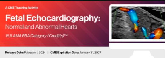
2024 Fetal Echocardiography: Normal and Abnormal Hearts – A Video CME Teaching Activity
$45.00
This Product is shared via google drive download link, So please share your correct Gmail id while placing the order .Please note that there are no CME points or certificate associated with this course Samples for Courses Can be found here : Free Samples Here!
2024 Fetal Echocardiography: Normal and Abnormal Hearts
Advanced Prenatal Cardiac Imaging and Congenital Heart Disease CME Program
Introduction
Fetal echocardiography has evolved into one of the most technically demanding and clinically important areas in prenatal imaging. As ultrasound technology advances and prenatal cardiac screening becomes increasingly sophisticated, clinicians are expected not only to recognize major structural abnormalities, but also to interpret subtle physiologic changes that may significantly alter perinatal management and neonatal outcomes.
2024 Fetal Echocardiography: Normal and Abnormal Hearts offers a detailed, clinically grounded exploration of modern fetal cardiac imaging, combining foundational anatomy with advanced diagnostic interpretation across a broad spectrum of congenital heart disease.
Rather than limiting the discussion to isolated pathology review, the activity approaches fetal echocardiography as a dynamic clinical process involving image optimization, Doppler assessment, physiologic interpretation, prenatal counseling, genetic considerations, and multidisciplinary care planning.
The result is a far more realistic educational experience for clinicians who routinely encounter fetal cardiac screening in practice.
Clinical Relevance
Prenatal diagnosis of congenital heart disease continues to play an increasingly important role in maternal-fetal medicine and pediatric cardiology. Early identification of cardiac abnormalities can significantly influence delivery planning, neonatal intervention strategies, and long-term outcomes for children born with congenital heart disease.
In practice, however, fetal cardiac imaging remains challenging.
Many sonographers and physicians encounter difficulty when distinguishing between normal developmental variation and subtle pathologic findings, particularly during early gestation or in technically limited examinations. Small deviations in outflow tract anatomy, venous return, rhythm disturbances, or ventricular symmetry may carry substantial clinical implications.
This CME activity addresses those challenges directly through detailed imaging review, real case interpretation, and extensive discussion of both normal and abnormal fetal cardiac anatomy.
Particular emphasis is placed on:
- cardiac chamber abnormalities
- outflow tract anomalies
- venous malformations
- fetal arrhythmias
- early gestation cardiac imaging
- Doppler interpretation
- image optimization strategies
- prenatal functional assessment
The educational structure mirrors real clinical workflow rather than isolated textbook categorization, which makes the material especially practical for clinicians actively performing or interpreting fetal cardiac examinations.
Educational Approach
Building Diagnostic Confidence Through Imaging Interpretation
One of the strongest aspects of this activity is its balance between foundational principles and advanced diagnostic reasoning.
The course begins with core concepts such as:
- national fetal echocardiography guidelines
- normal fetal cardiac anatomy
- screening protocols
- ultrasound optimization
- Doppler application
- first- and second-trimester cardiac assessment
From there, the curriculum gradually progresses into increasingly complex congenital pathology and physiologic abnormalities.
Importantly, the lectures do not simply label defects. Faculty frequently discuss:
- how abnormalities present sonographically
- common diagnostic pitfalls
- image acquisition challenges
- differential considerations
- clinical implications of missed findings
- practical scanning strategies
In fetal echocardiography, subtle image interpretation errors can significantly alter downstream care. That nuance is reflected throughout the course.
The inclusion of “Just Images & Movie Clips” sessions is particularly valuable because pattern recognition remains one of the most difficult skills to teach in fetal cardiac imaging.
Key Learning Areas
Normal Fetal Cardiac Anatomy & Screening
A substantial portion of the program focuses on establishing a strong understanding of normal anatomy before progressing into pathology.
Topics include:
- cardiac chambers
- great vessels
- upper mediastinum
- venous anatomy
- fetal circulation
- first-trimester screening
- second-trimester cardiac evaluation
The discussions surrounding standardized screening protocols are especially useful for clinicians seeking to improve consistency and detection rates in routine obstetric imaging.
PROGRAM
- National Guidelines for Fetal Echocardiography: What is Included?
Tracy L. Anton, BS, RDMS, RDCS, FAIUM - Normal Fetal Cardiac Anatomy: The Cardiac Chambers
Julia E. Solomon, MDCM, FACOG, FAIUM - How to Screen for Congenital Heart Disease in the First & Second Trimester
Elena Sinkovskaya, M.D., Ph.D., RDMS, RDCS - Optimizing Your Image in Fetal Cardiac Screening
Tracy L. Anton, BS, RDMS, RDCS, FAIUM - Just Images & Movie Clips: Do You Know the Diagnosis?
Elena Sinkovskaya, M.D., Ph.D., RDMS, RDCS - Anomalies of the Cardiac Chambers I: Atrial & Ventricular Septal Defects
Julia E. Solomon, MDCM, FACOG, FAIUM - Anomalies of the Cardiac Chambers 2: Atrioventricular Septal Defects
Edgar Jaeggi, M.D., FRCPC - Anomalies of the Cardiac Chambers 3: Hypoplastic Left Heart Syndrome & Critical Aortic Stenosis
Anita J. Moon-Grady, M.D. - Anomalies of the Cardiac Chambers 4: Abnormal Right Ventricle (Tricuspid Atresia, Ebstein Anomaly, Pulmonary Atresia with Intact Septum)
Bettina F. Cuneo, M.D. - Hands-on Scanning Session 1: Image Optimization for Cardiac Screening
Tracy L. Anton, BS, RDMS, RDCS, FAIUM - Anomalies of the Cardiac Chambers 5: Single Ventricle-Type Congenital Heart Disease
Bettina F. Cuneo, M.D. - Systemic Fetal Venous Malformations: A Standardized Approach to Diagnosis
Alfred Abuhamad, M.D. - Fetal Ectopy and Tachyarrhythmias: Diagnosis & Management
Edgar Jaeggi, M.D., FRCPC - Case Presentation I
Julia E. Solomon, MDCM, FACOG, FAIUM - Case Presentation II
Edgar Jaeggi, M.D., FRCPC - Artificial Intelligence in Fetal Cardiac Imaging
Anita J. Moon-Grady, M.D. - Normal Fetal Cardiac Anatomy: The Great Vessels & Upper Mediastinum
Alfred Abuhamad, M.D. - Tips & Tricks of Fetal Echocardiography
Tracy L. Anton, BS, RDMS, RDCS, FAIUM - Genetic Aspects of Congenital Heart Disease
Julia E. Solomon, MDCM, FACOG, FAIUM - Just Images & Movie Clips: Do You Know the Diagnosis?
Elena Sinkovskaya, M.D., Ph.D., RDMS, RDCS - The Use of 3D/4D Ultrasound in Fetal Cardiac Imaging
Elena Sinkovskaya, M.D., Ph.D., RDMS, RDCS - Anomalies of Great Vessels 1: Transposition of Great Vessels
Julia E. Solomon, MDCM, FACOG, FAIUM - Anomalies of the Great Vessels 2: Tetralogy of Fallot
Alfred Abuhamad, M.D. - Anomalies of the Great Vessels 3: Aortic Arch Abnormalities
Anita J. Moon-Grady, M.D. - Hands on Scanning Session 2: The Cardiac Exam-Gray Scale and Color Doppler
Alfred Abuhamad, M.D. - Bradyarrhythmias and Long QT Syndrome
Bettina F. Cuneo, M.D. - Anomalies of Pulmonary Venous Return
Anita J. Moon-Grady, M.D. - Fetal Cardiac Interventions
Edgar Jaeggi, M.D., FRCPC - Case Presentation III
Elena Sinkovskaya, M.D., Ph.D., RDMS, RDCS - Hands on Scanning Session 3: Doppler / Arrythmias
Elena Sinkovskaya, M.D., Ph.D., RDMS, RDCS & Faculty - Update on Management of Pregnancies with Sjogren’s Antibodies
Bettina F. Cuneo, M.D. - Cardiac Imaging in Early Gestation
Alfred Abuhamad, M.D. - Prenatal Evaluation of Cardiac Function
Elena Sinkovskaya, M.D., Ph.D., RDMS, RDCS - Heterotaxy Syndrome
Edgar Jaeggi, M.D., FRCPC - Review of Course: Fetal Cardiac Imaging Know What to Look For
Alfred Abuhamad, M.D. - Just Cases
Elena Sinkovskaya, M.D., Ph.D., RDMS, RDCS and Tracy L. Anton, BS, RDMS, RDCS, FAIUM
Related products

Neurology / neurosurgery courses

Critical Care - Emergency medicine
The Brigham Board Review in Infectious Diseases Brigham and Women’s Hospital Board Review

Critical Care - Emergency medicine
Anesthesiology & pain medicine
Critical Care - Emergency medicine
Gulfcoast Ultrasound-Guided Upper Extremity Nerve Blocks for Emergency Medicine

Critical Care - Emergency medicine